


The exams
The coronography
What is a coronography ?
The exam by coronarography consists in a filmed radiography of your coronary arteries. It
also includes a sequence evaluating the correct functioning of the left ventricle. A coronarography can detect one or more spots of shrinking which are the origin of pains or aninfarct, therefore making a better adapted treatment possible.
This treatment is using X-rays and a tracer product based on iodine. Its aim is to permit to view the coronary arteries, irrigating the heart. Into the vessel is introduced a catheter in order to inject the tracer product, which mixes with the blood: the X-ray images will show the vascular system under the influence of the radio-opaque specification of the iodine.
.
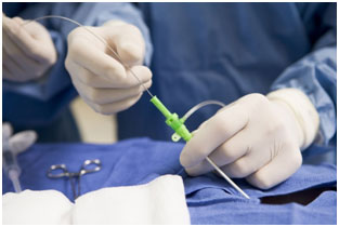
How is the coronography being carried out ?
A cardiologist is carrying out this exam. A venous perfusion will be placed on your arm. You will be lying on your back during the exam. Permanently, your cardiac rhythm and your blood pressure is recorded. The exam is made under sterile conditions and with local anaesthetic. The cardiologist will disinfest your skin, then applies the local anaesthetic. Once the specific zone anaesthetized, the he will introduce a catheter in an artery of the fold of the hip or the arm. He will lead the catheter to the beginning of the coronary arteries, passing by the aorta. Once in place, the tracing product is injected and then several images will be made to make the vascular network visible.
The exam will last approximatively between 30 minutes and one hour. After the exam the catheter is taken out and the vessel will be compressed, thus stopping any bleeding.